

3 Eye Centers in South Central Ohio to Serve You Better
With offices in Chillicothe, Greenfield and Waverly, Chester Eye Center serves South Central Ohio with the latest technology in eye care. Our eye care offices use cutting-edge digital imaging technology to assess your eyes. Many eye diseases, if detected at an early stage, can be treated successfully without total loss of vision. Digital imaging and other specialized testing can be very important in assisting your Chillicothe optometrist to detect and measure any subtle changes in the health of your eyes. Many eye conditions, such as glaucoma, diabetic retinopathy and macular degeneration are diagnosed by detecting changes over time.
Cirrus Optical Coherence Tomography (OCT)
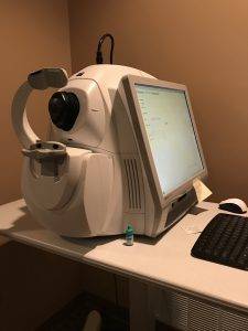 Cirrus HD-OCT enables your optometrist to perform multiple analyses for glaucoma.
The three most common tests are:
- Retinal Nerve Fibre Layer (RNFL) Analysis reveals the thickness of the layer of the retina that contains nerve fibres that travel up the optic nerve. If glaucoma is present, that layer may gradually lose thickness.
- Macular Thickness Analysis examines the condition and thickness of the macula, which is the part of the retina that provides central vision. Thinning of the macula is a possible sign of glaucoma progression.
- Optic Nerve Head Analysis reveals the structure of the optic nerve where it originates in the retina. With glaucoma, the “cup” in the optic nerve may enlarge.
Humphrey Visual Field Testing
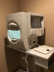 A visual field test measures the range of your peripheral or “side” vision to assess whether you have any blind spots (scotomas), peripheral vision loss or visual field abnormalities. It is a straightforward and painless test that does not involve eye drops but does involve the patient’s ability to understand and follow instructions.
An initial visual field screening can be carried out by the optometrist by asking you to keep your gaze fixed on a central object, covering one eye and having you describe what you see at the periphery of your field of view. For a more comprehensive assessment, special equipment might be used to test your visual field. In one such test, you place your chin on a chin rest and look ahead. Lights are flashed on, and you have to press a button whenever you see the light. The lights are bright or dim at different stages of the test. Some of the flashes are purely to check you are concentrating. Each eye is tested separately and the entire test takes 15-45 minutes. These machines can create a computerized map out your visual field to identify if and where you have any deficiencies.
Digital Fundas Camera
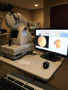 This equipment takes a picture of the retina,often without dilation drops. It can also be used as a wide-angled anterior segment camera to take pictures of eye lids, lashes, subconjunctival hemorrhages, etc. We use it for routine eye exams, diabetes, Hypertension glaucoma, retinopathy (retinal disease) and high risk patients.
TearLab Technology
Tear osmolarity has been shown to have the best predictive value for diagnosing Dry Eye Disease of any single test. TearLab’s sophisticated lab on a chip technology tests a tiny tear sample to measure Osmolarity, or the salt content in the tears. The Doctor will test both eyes, and will take the highest number of the two tests, to generate Your Osmolarity Number. Your Osmolarity Number gives doctors a meaningful measure of the health and stability of the protective tear film that covers the surface of your eyes. The number generated correlates with the severity of the disease.
Equipped with the TearLab Osomolarity System, our optometrists can now use quantitative data at the point of care to aid in the diagnosis of DED and to improve our ability to manage these patients.
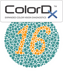 ColorDx
ColorDx: A detailed color vision evaluation that determines if there is loss of color perception as a result of optic nerve head disease, diabetic eye disease or other neurological eye disease. Tritan deficiencies (blue color vision defects) are rarely genetic, yet as high as 15% of the general population may have a tritan deficiency. This is an important but often overlooked sign that may be caused by retinal, optic nerve, or neurological disorders, in addition to trauma, cataracts, and hundreds of common drugs and substances.
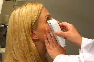
B-Scan Ophthalmic Ultrasound
 B-scan ultrasonography is an important adjuvant for the clinical assessment of various ocular and orbital diseases.
The ultrasound uses high-frequency sound waves that travel through the eye. Reflections (echoes) of the sound waves form a picture of the structure of the eye.
Your eye is numbed anesthetic eye drops. The ultrasound probe is placed against the front surface of the eye. The test takes about 15 minutes. You will be seated and you may be asked to look in many directions. The test is usually done with your eyes closed. A gel is placed on the skin of your eyelids. The B-scan probe is gently placed against your eyelids to do the test.
B-scan is done to look at the inside part of the eye or the space behind the eye that can’t be seen directly. This may occur when you have cataracts or other conditions that make it hard for the doctor to see into the back of your eye. The test may help diagnose retinal detachment, tumors, or other disorders.